Lista 103+ Foto mosaico fluido de la membrana celular El último
¡Descubre las increíbles imágenes de mosaico fluido de la membrana celular en huanluyenantoan.edu.vn! Este sitio web ha recopilado cuidadosamente y elaborado una selección de imágenes. Además, hay más imágenes relacionadas disponibles en mosaico fluido de la membrana celular . ¡No te lo pierdas!
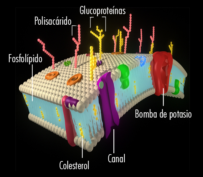
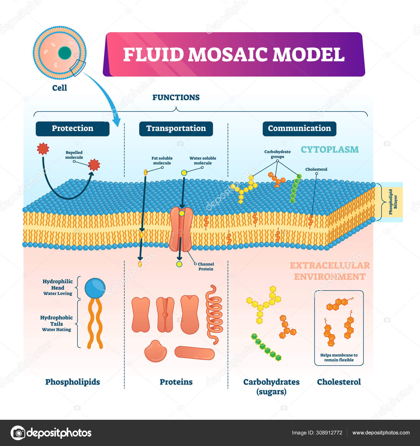
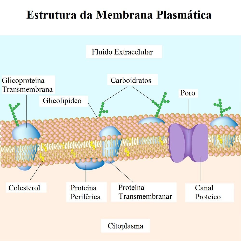
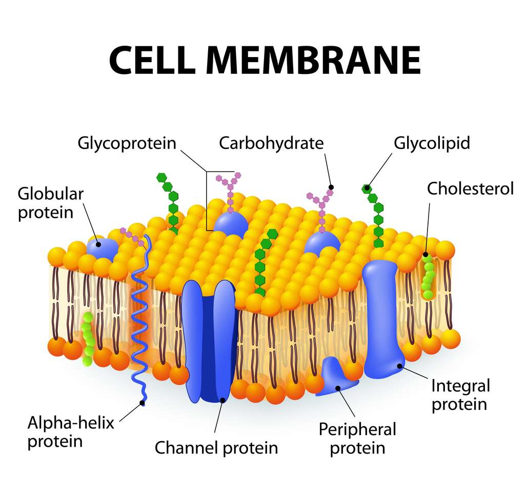
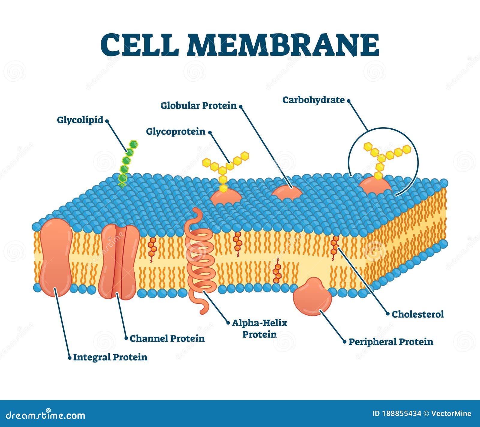
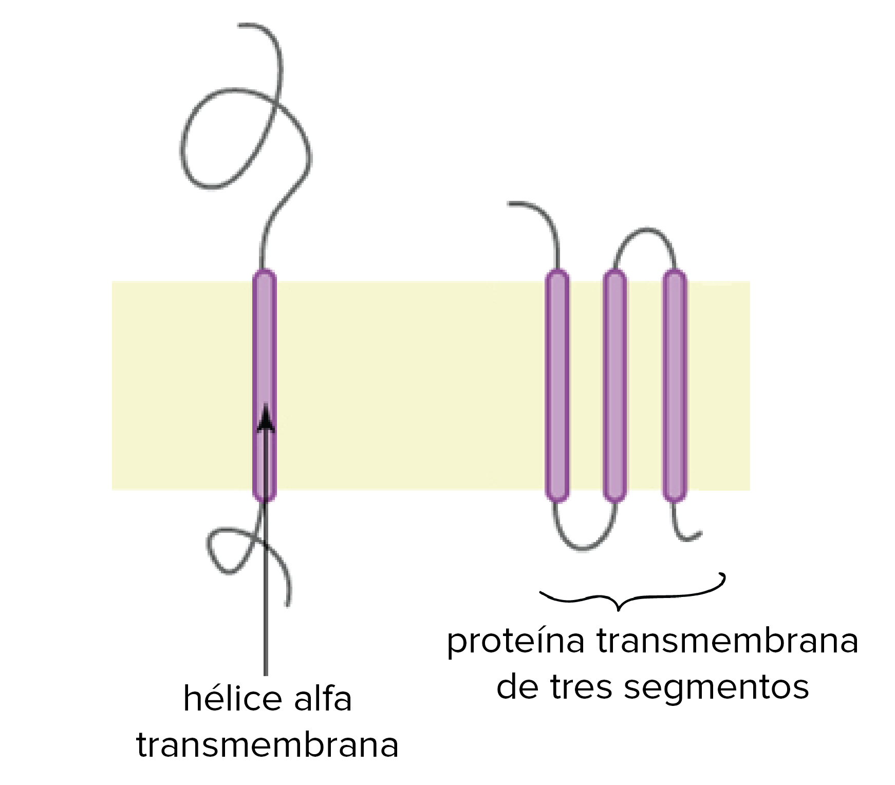
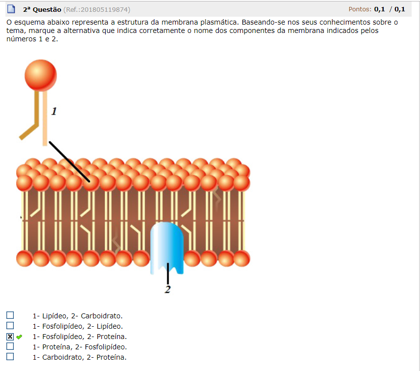
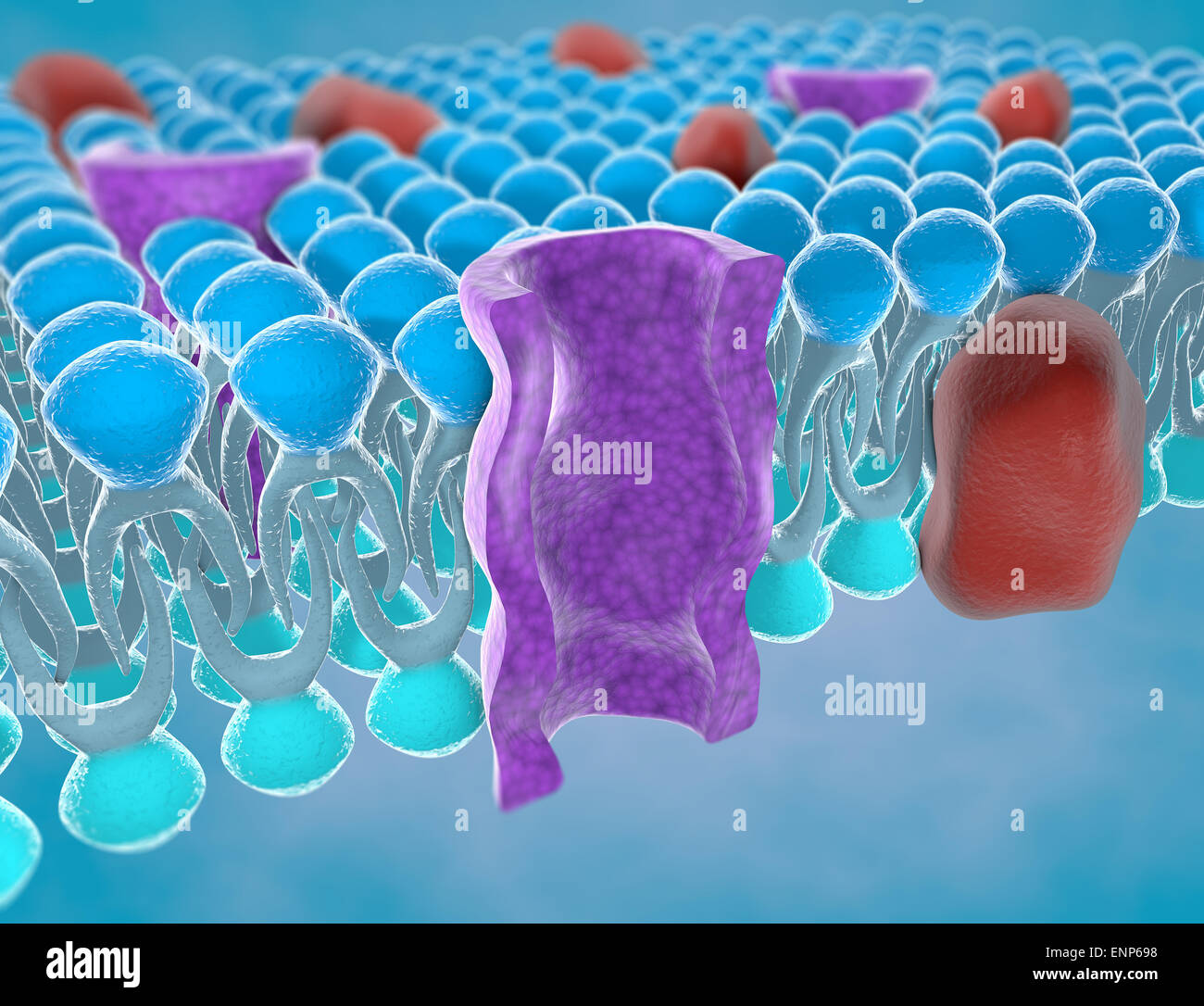
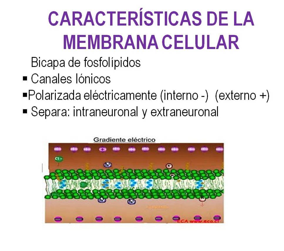
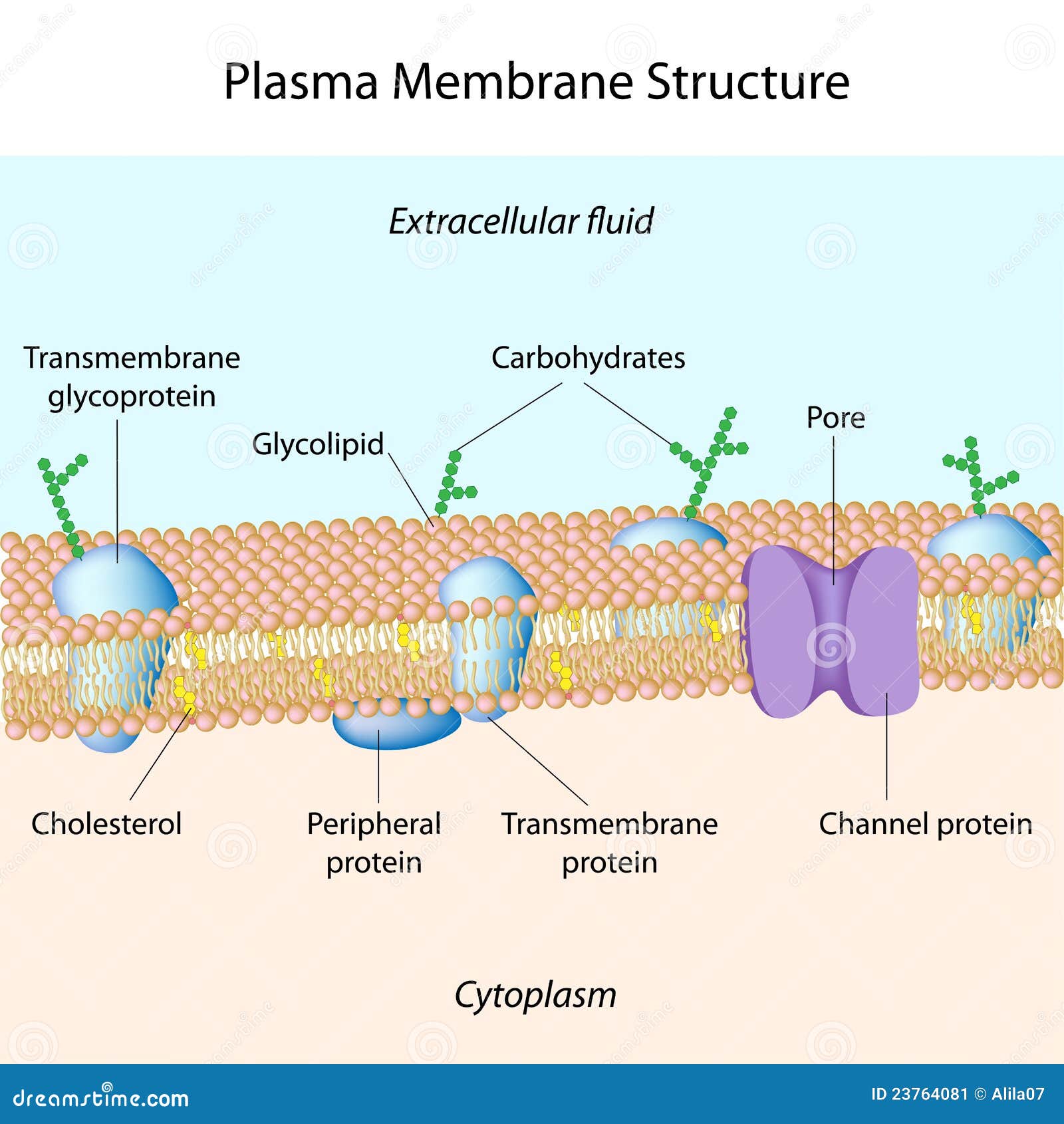
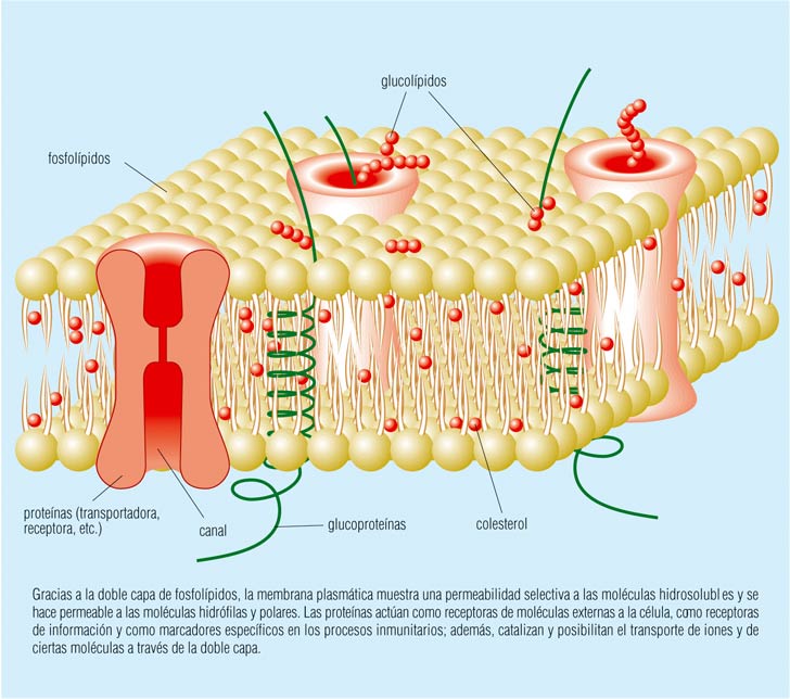
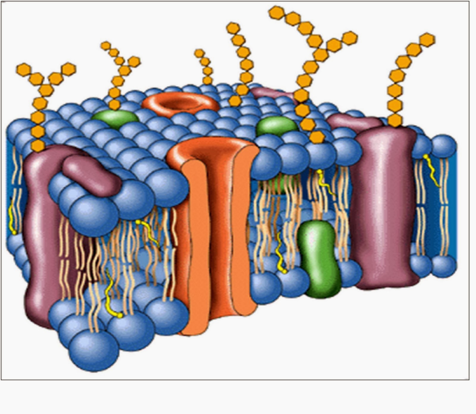
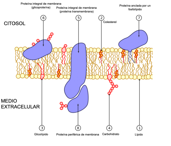
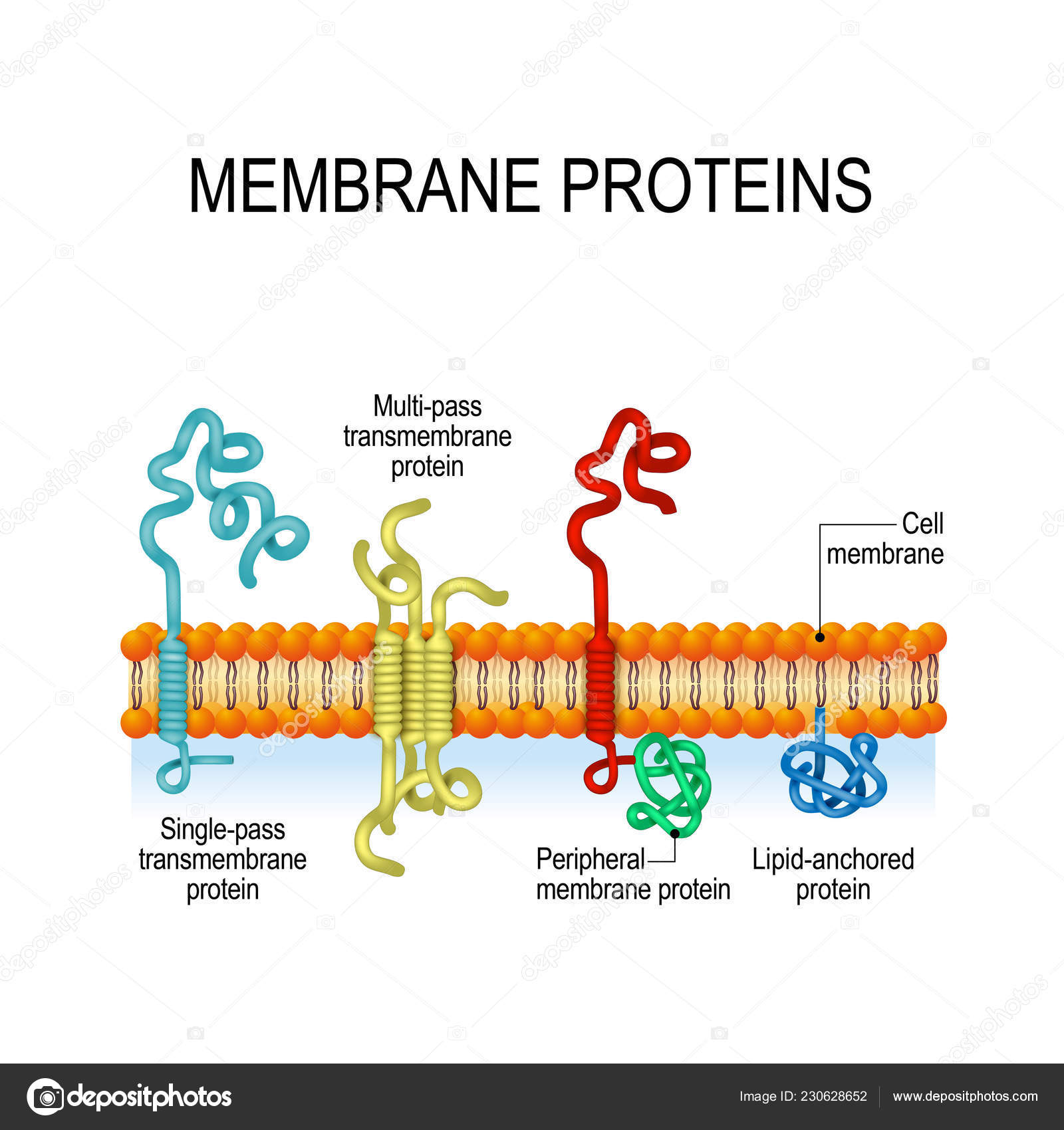
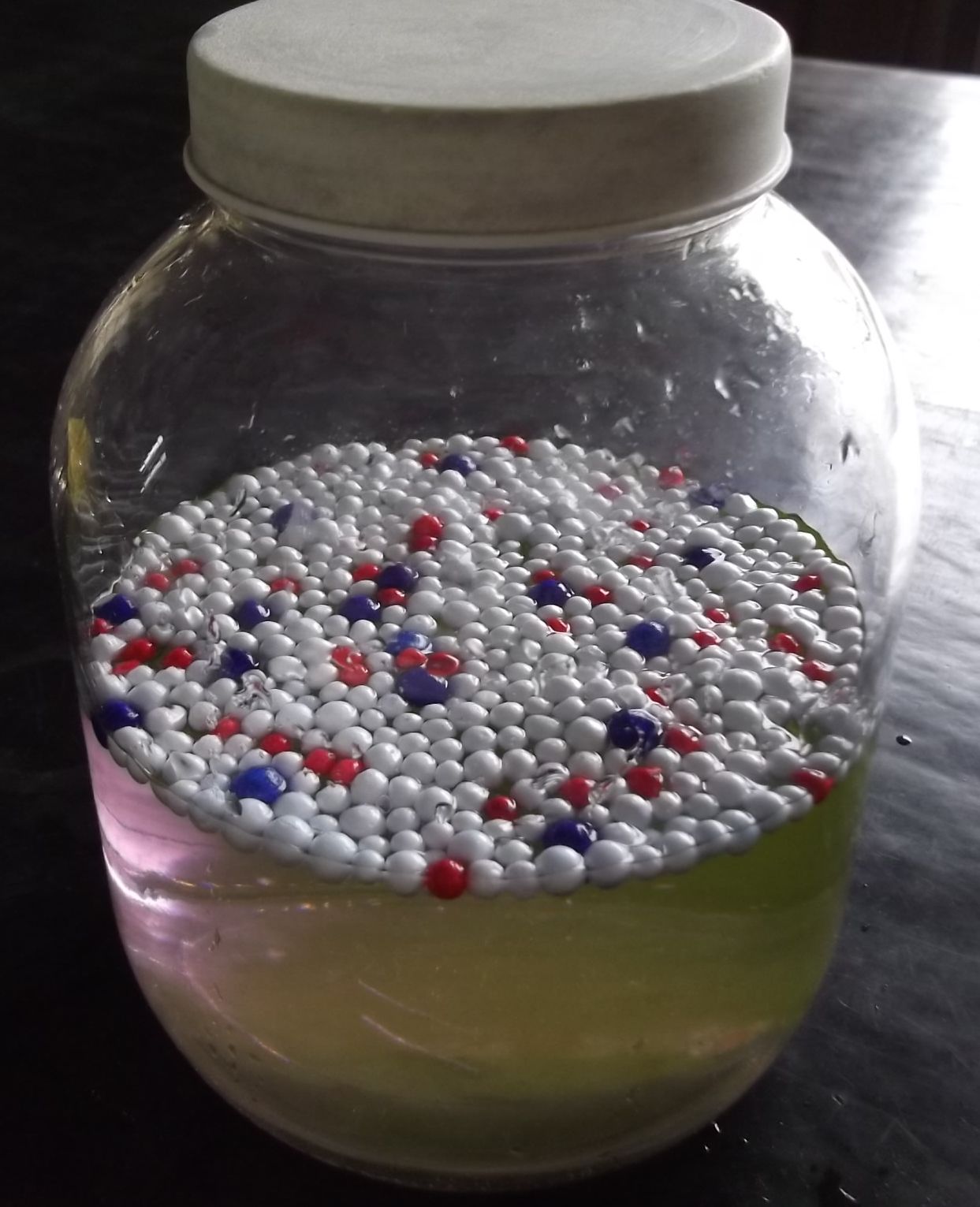
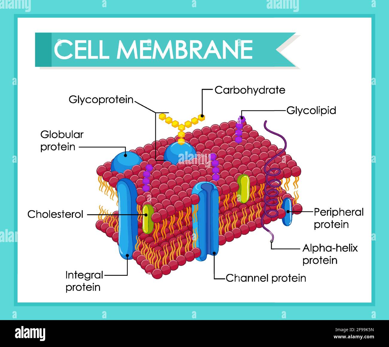
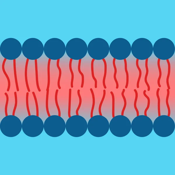
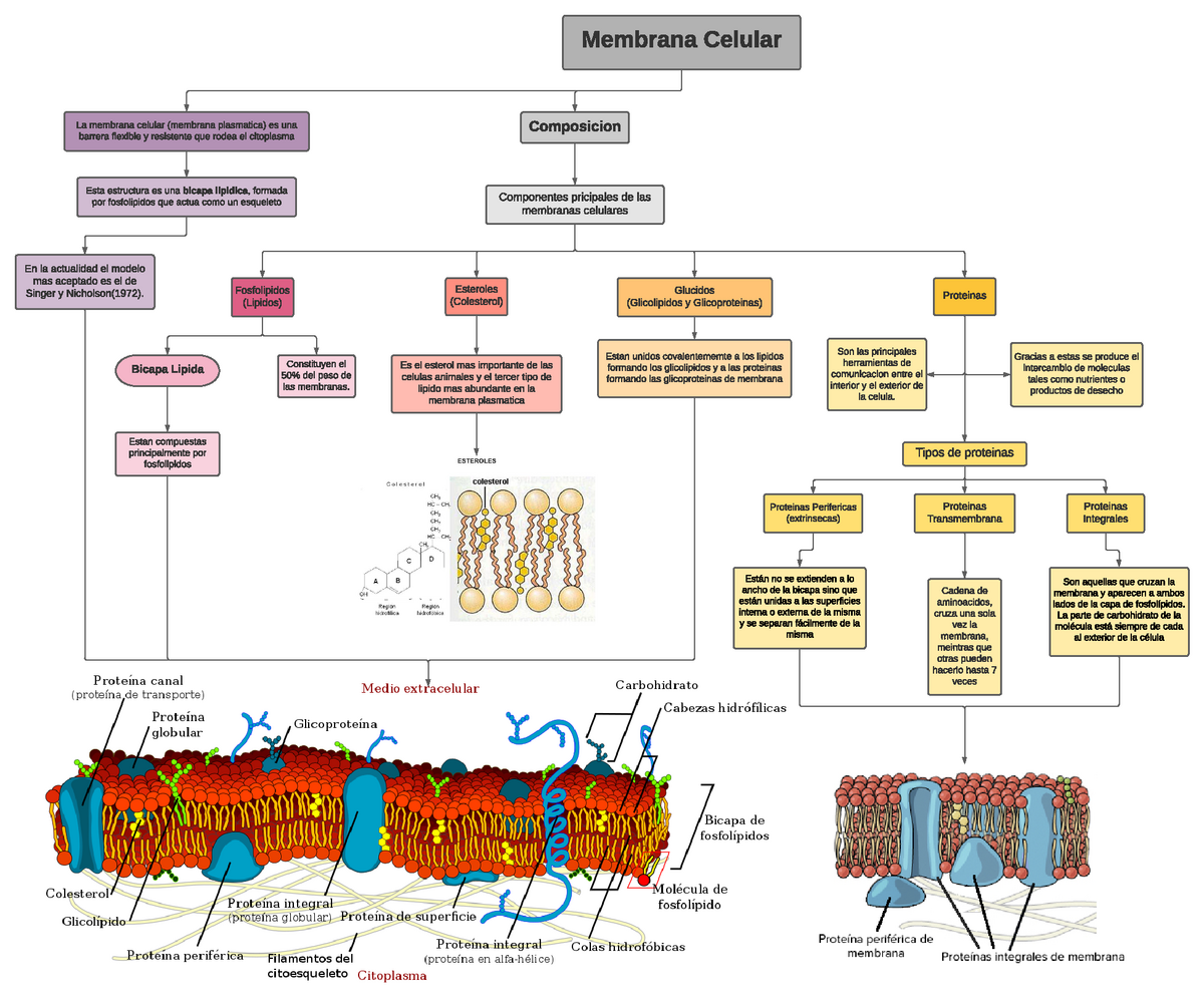
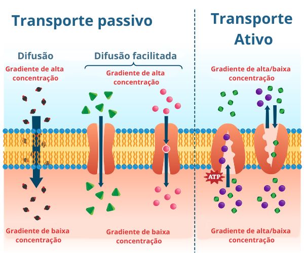
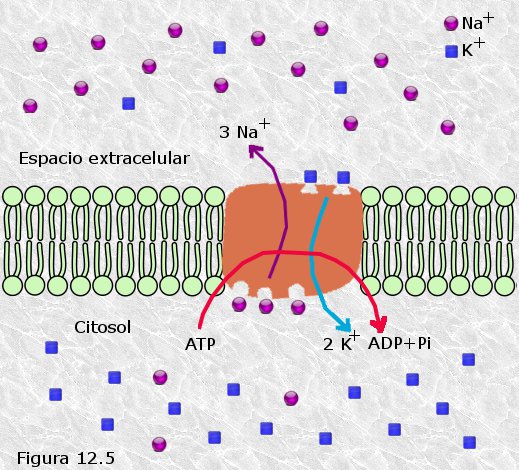
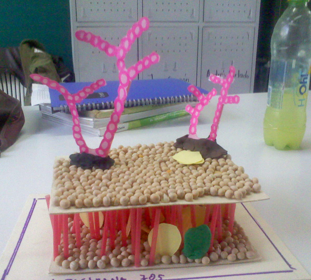
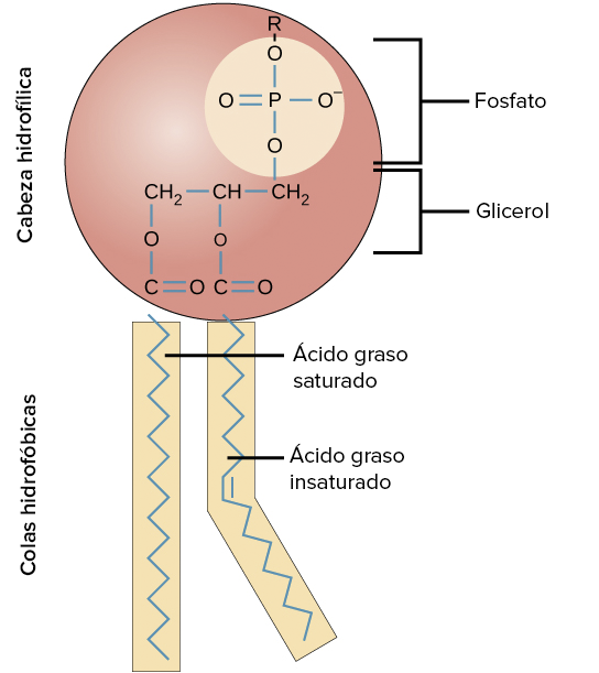
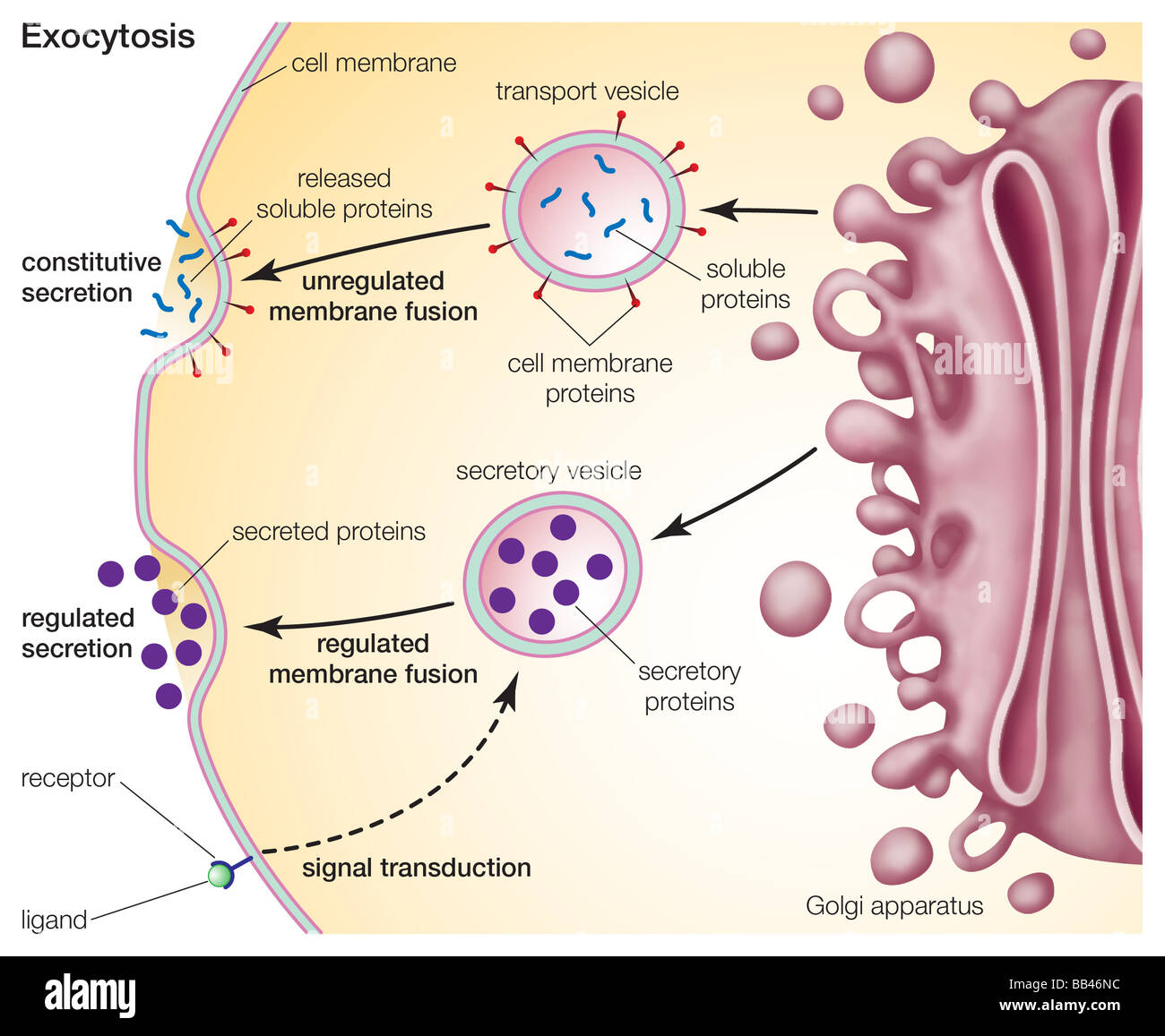
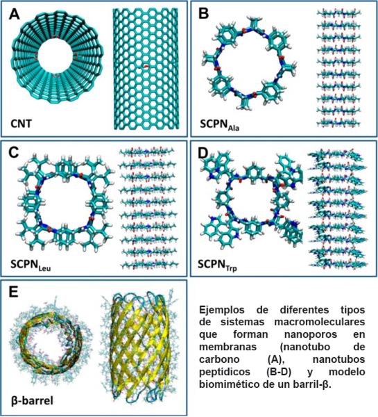
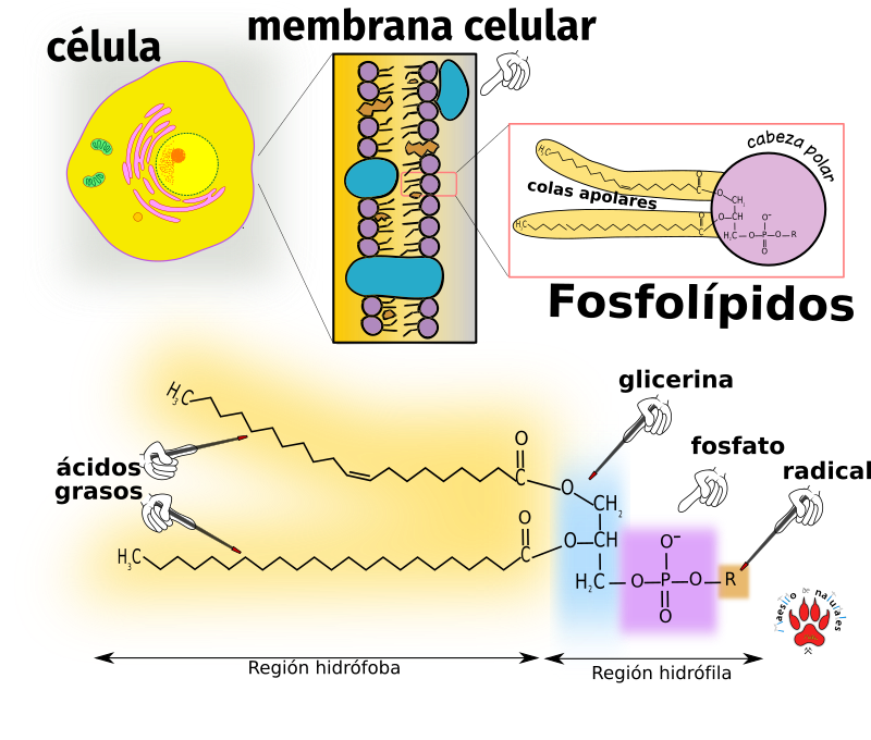
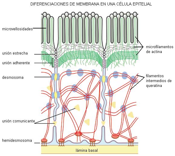
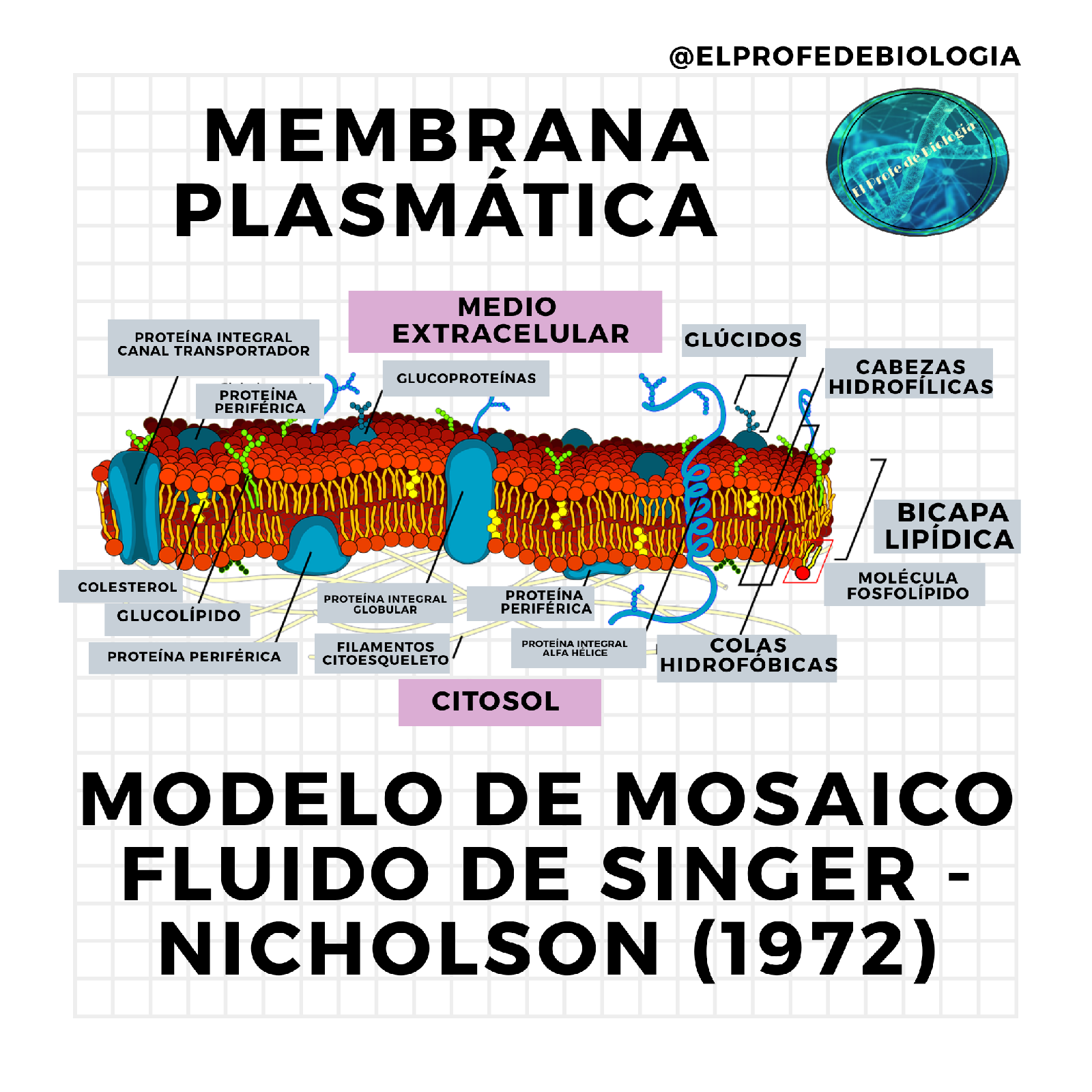
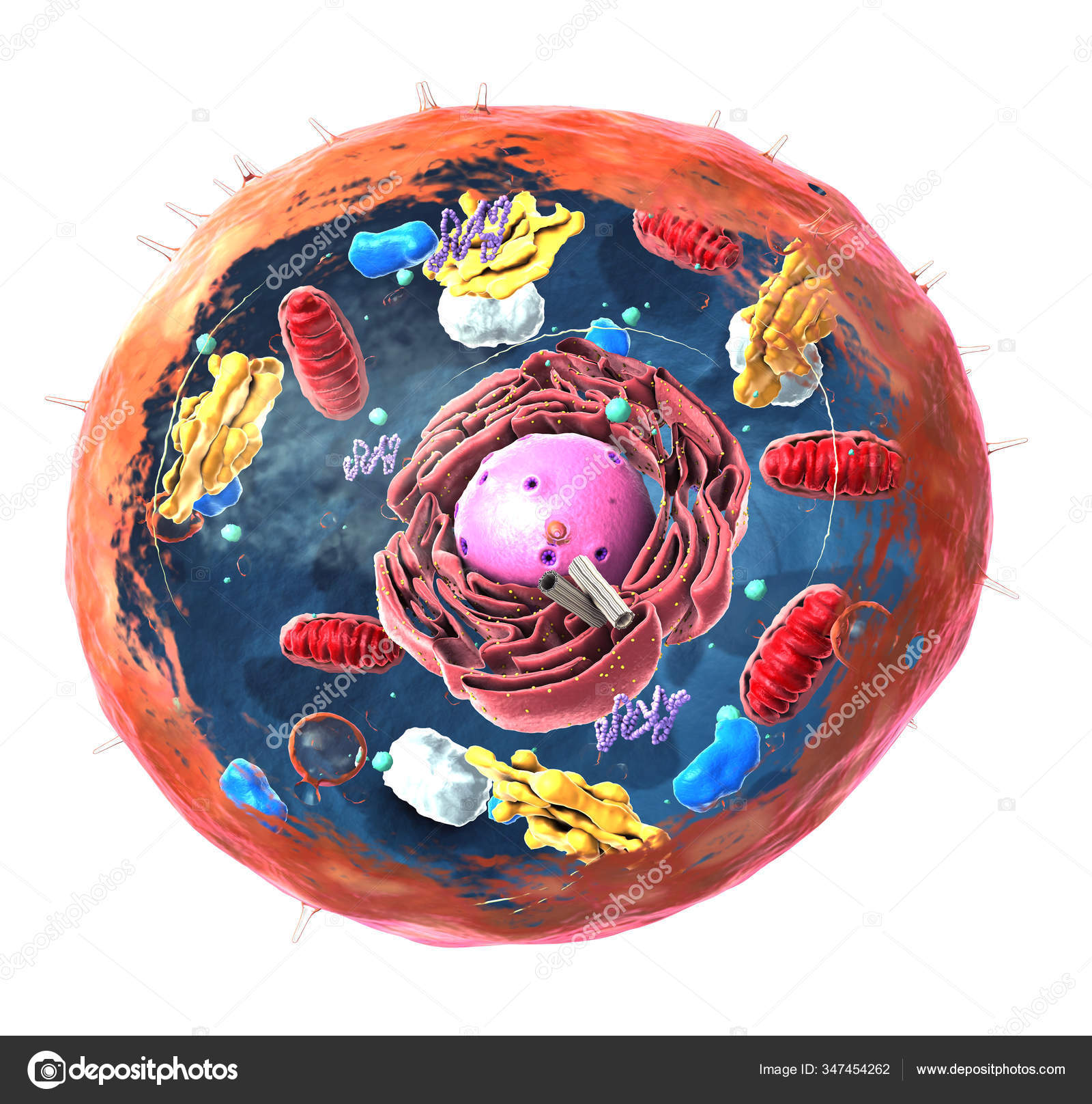
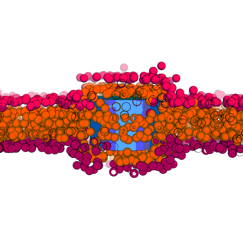
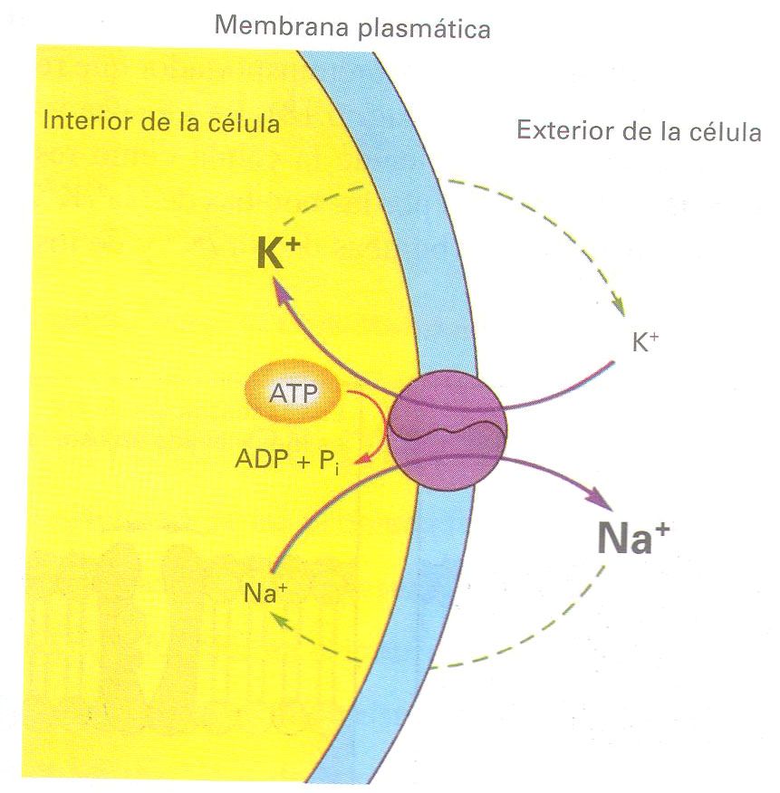
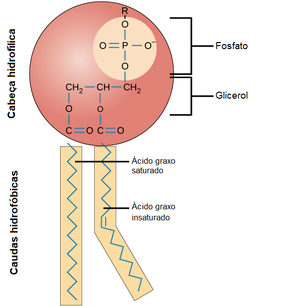
mosaico fluido de la membrana celular


¡Agradecemos que hayas leído el apasionante artículo sobre mosaico fluido de la membrana celular en huanluyenantoan.edu.vn! No dudes en comentar y descubrir más artículos relacionados en la sección siguiente. Esperamos que encuentres información valiosa e interesante.
Posts: mosaico fluido de la membrana celular
Categories: Sintetizar imágenes
Author: huanluyenantoan.edu.vn